
Measurements and Results
W.H. Zachariasen first proposed that amorphous silica has a random network structure (1). Crystalline silica is composed only of 6-membered rings of Si atoms, while amorphous silica is composed of n-membered rings as shown in Fig. 1. To determine the network structure, RMC simulation was performed using RMCProfile software (2). The simulation was started with 1500 Si and 1500 O atoms generated in a 35Å3 cell, taking into account the atomic density. The simulation results showed good agreement between the observed and simulated PDF patterns (Fig. 2). The network structure was obtained from the RMC simulation (Fig. 3) and the population of n-membered rings was derived using the ISAACS software (3). As shown in Fig. 4, 6-membered rings had the highest frequency, and there were n-membered rings that indicate that amorphous silica has the proposed network structure (4).
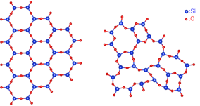
Fig. 1: The structures of crystalline silica (left) and amorphous silica (right)
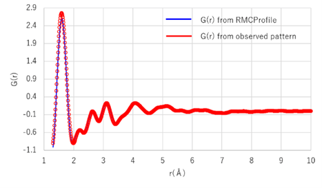
Fig. 2: Observed and simulated PDF patterns
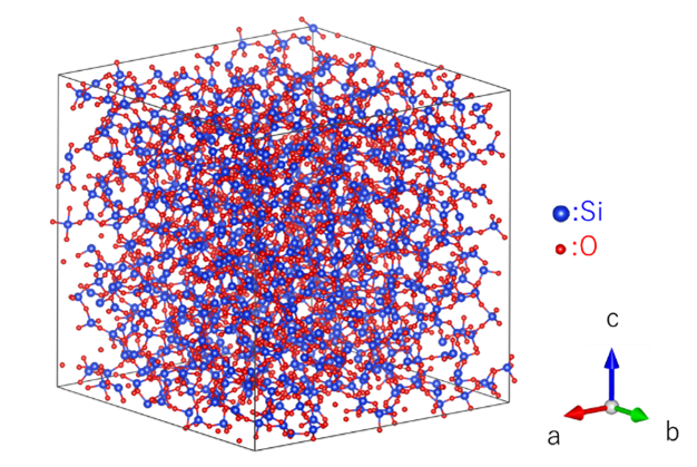
Fig. 3: amorphous silica structure obtained from RMC
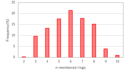
Fig. 4: n-membered ring frequency of Si-Si
References: (1) R. L. McGreevy et al.: Molecular Simulation, 1 (1988) 359-367.
(2) W. H. Zachariasen: J.Am.Chem. Soc., 54 (1932) 3841-3851.
(3) S.Le Roux et al.: J.Appl.Cryst., 43 (2010) 181-185.
(4) S.Kohara et al.: Proc. Nat.Acad.Sci., 108 (2011) 14780-14785.
Recommended equipment and software
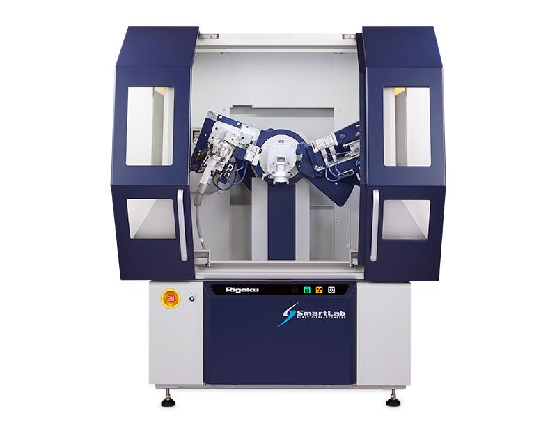
► Automated multipurpose X-ray diffractometer SmartLab + High-resolution and high-speed 1D detector
D/tex Ultra250 HE + Ellipsoidal multilayer X-ray mirror CBO-E
► Integrated X-ray analysis software SmartLab Studio II (PDF plugin)
RIGAKU RECOMMENDS
SmartLab